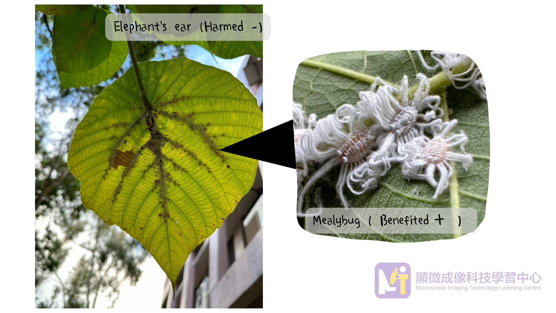
Recently, I had a lesson with my students learning the symbiotic relationships among organisms. We observed some mealybugs on the leaves of the Elephant's Ear (Macaranga tanarius) and they were sucking sap on the leaf veins. Though the plant would not die, the mealybugs were benefited at the expense of the plant. It is a typical parasitism.
Fig 1. Parasitism between Mealybug and Elephant’s ear
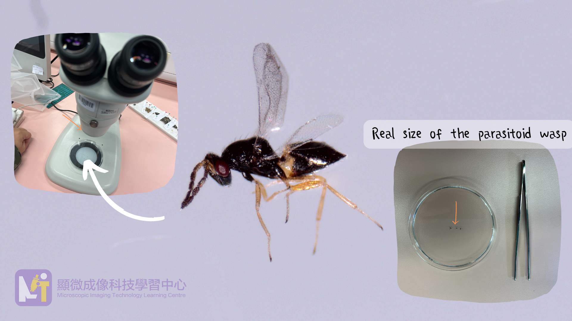
A student found that one of the mealybugs was in black color instead of white, thus we decided to remove its waxy secretion to find the reason. Unexpectedly, there were many tiny‘black dots’ flying out from the waxy secretion. We observed one of these “dots”under the optical stereo-microscope, and found that it was actually a kind of parasitoid wasp!
For the external morphology of this parasitoid wasp, it has black body, red compound eyes, yellowish legs, hidden ovipositor and a pair of transparent wings.
Fig 2. Observation under optical stereo microscope
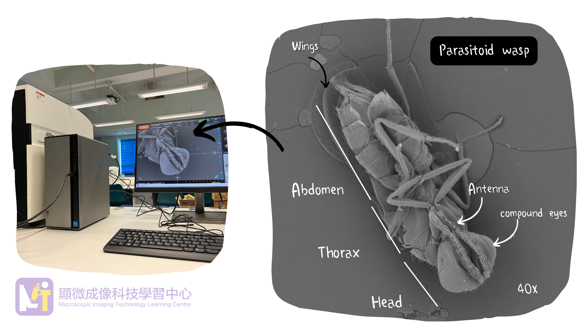
This parasitoid wasp was too small so we decided to put it into the scanning electron microscope for a detailed observation. We could see more microstructures of the insect after magnifying it in 40 times. The resolution and magnification power of the scanning electron microscope are much higher than the optical stereo-microscope, allowing us to see more details of the parasitoid wasp.
Fig 3. Observation under scanning electron microscope
This parasitoid wasp was too small so we decided to put it into the scanning electron microscope for a detailed observation. We could see more microstructures of the insect after magnifying it in 40 times. The resolution and magnification power of the scanning electron microscope are much higher than the optical stereo-microscope, allowing us to see more details of the parasitoid wasp.
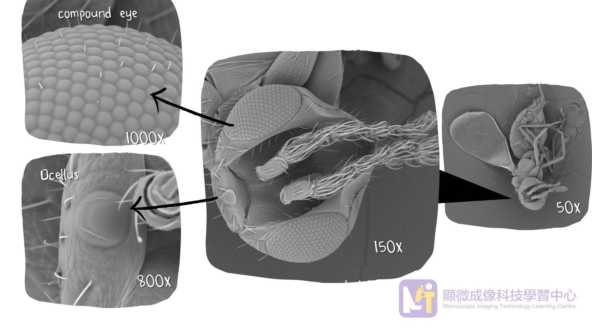
Fig 4. Parasitoid wasp's head (eyes) under scanning electron microscope
We started our observation from the parasitoid wasp’s head, it has two large compound eyes. When we magnified it to 1000 times, we could see its compound eyes were composed of many ommatidia with hairs in between. In addition, there was an ocellus, for light detection, between the two compound eyes.
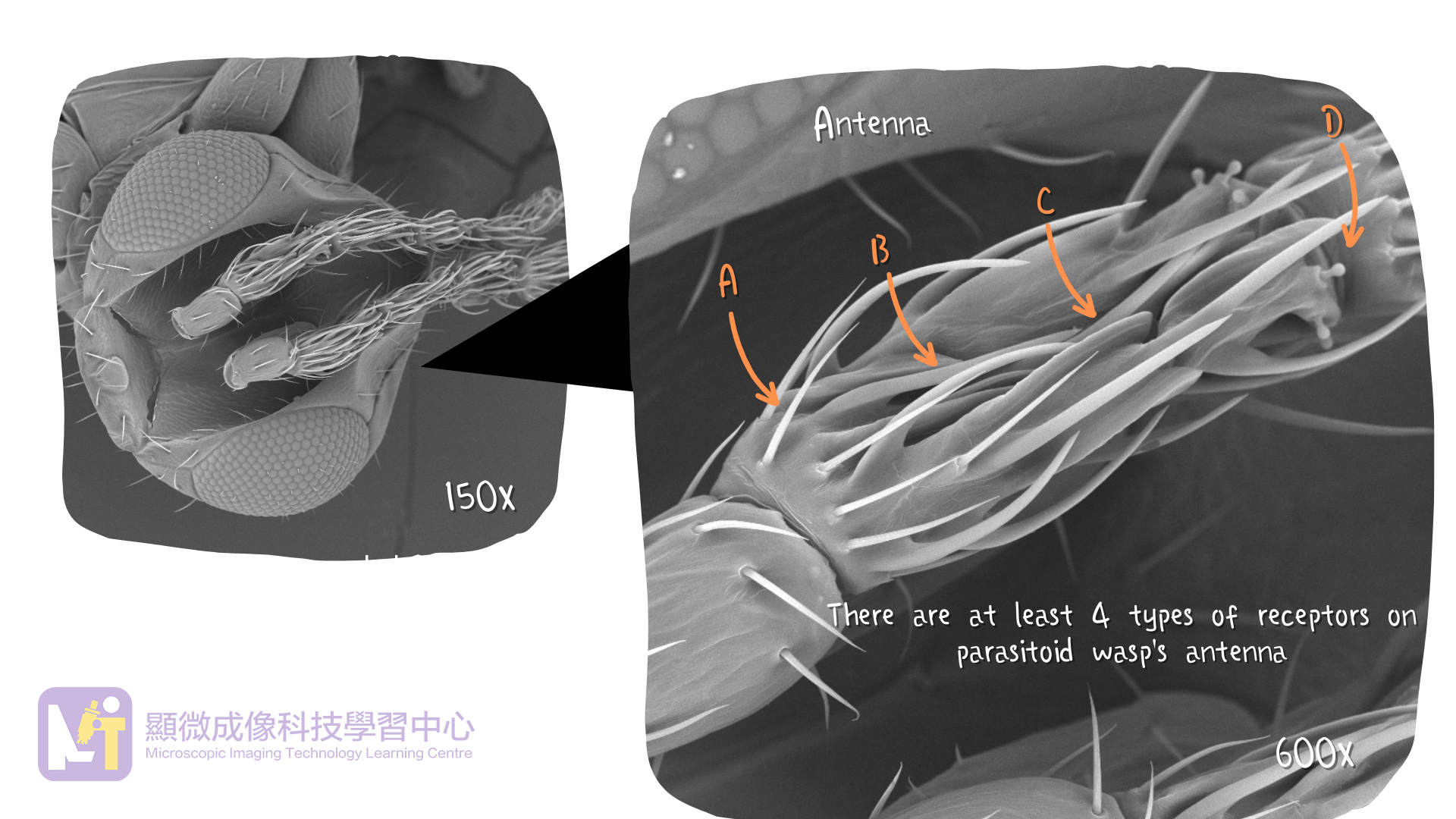
When we magnified the parasitoid wasp to 600 times, we took a closer look to its antenna and found at least four different types of receptors on it, including:
Receptor A : hair-like receptor grown from the antenna
Receptor B : hair-like receptor on the antenna
Receptor C : tabular receptor on the antenna
Receptor D : papillary-like receptor on the antenna
Different receptors are used to receive different external stimuli. With more receptors, the insect could receive more types of stimuli. Some studies (in-text citation needed) have pointed out that the antennae of parasitoid wasps of the same species have sexual dimorphism. Some of the receptors are more developed in the males for finding mates, while these kinds of receptors are weaker in the females. In contrast, female needs stronger sensation to find hosts for laying eggs, their respective receptors on finding hosts are stronger than the males’.
Fig 5. Parasitoid wasp's head (Antenna)under scanning electron microscope
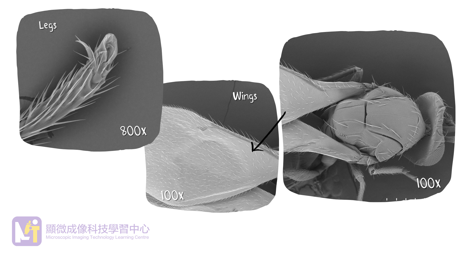
Three pairs of legs and two pairs of wings were extended from the parasitoid wasp's thorax. These body parts had fine hairs on the surface. There were small claws at the end of the legs.
Fig 6. Parasitoid wasp's thorax under scanning electron microscope
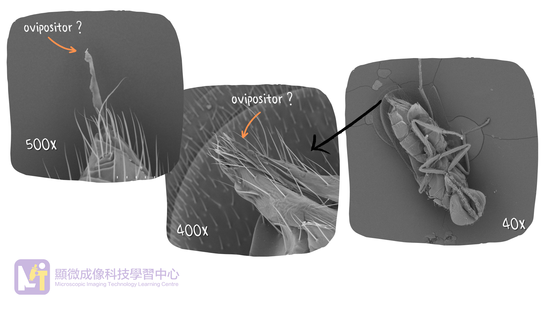
There was a long structure that looked like an ovipositor at the end of the abdomen.
Fig 7. Parasitoid wasp' abdomen under scanning electron microscope
The female parasitiod wasp lays eggs on the mealybugs. Their larvae absorb the body fluid of the mealybugs for growing. When the body fluid of the mealybugs was all sucked and the mealybugs would die. The parasitiod wasp got benefits at the expense of the lives of the mealybugs. This is a host-parasitoid relationship.
The relationship among organisms in nature is intricate and delicately balanced.
Taking a look closely, you could also have special discoveries!
You might read more about Parasitoid-host interactions in Ho Koon Biology Page’s Instagram!
Sun Y. Y., Qin D. Y., Pan L., Mu Y. R., Yang Y. X., Xiang W. F., Zhu G. P. & Li. M.2020). Ultrastructure of Sexual Dimorphism in Antennal Sensilla of Endoparasitoid Chouioia cunea (Hymenoptera: Eulophidae). Scientia Silvae Sinicae, 56(10): 135-144. http://www.linyekexue.net/CN/abstract/abstract8680.shtml
Zhou H., Wu W.J., Zhang F. P. & Fu Y. G. (2013). Scanning Electron Microscopy Studies of the Antennal Sensilla of Metaphycus parasaissetiae Zhang & Huang (Hymenoptera: Encyrtidae). Neotropical Entomology, 42(3):278-87. https://www.researchgate.net/publication/255958103_Scanning_Electron_Microscopy_Studies_of_the_Antennal_Sensilla_of_Metaphycus_parasaissetiae_Zhang_Huang_Hymenoptera_Encyrtidae